Xstrahl in Action
Advances in conformal radiotherapy (RT) techniques and image-guidance have significantly reduced organs at risk (OAR) exposure, which has been associated with improved safety profiles. However, opportunities remain to further optimise treatments based on the preferential avoidance of discrete radiosensitive subregions of OARs that have functional significance.
Radiation induced cardiotoxicity (RICT) is an important sequela of radiotherapy to the thorax for patients. In this study, researchers aimed to investigate the dose and fractionation response of RICT. They propose global longitudinal strain (GLS) as an early indicator of RICT and investigate myocardial deformation following irradiation.
Publication
Dose-dependent changes in cardiac function, strain and remodelling in a pre-clinical model of heart base irradiation
Authors
Mihaela Ghita-Pettigrew, Kevin S. Edgar, Refik Kuburas, Kathryn H. Brown, Gerard M. Walls, Cecilia Facchi, David J. Grieve, Chris J. Watson, Alan McWilliam, Marcel van Herk, Kaye J. Williams, Karl T. Butterworth

Key Findings
- Functional features of radiation induced cardiotoxicity (RICT) show a biological effective dose (BED) dependence at 50 weeks after irradiation.
- Global longitudinal strain (GLS) shows changes observed as early as 10 weeks post base targeted irradiation, indicative of late loss of cardiac function.
- Segmental strain indicates the base of the heart as an early responding substructure as early as 10 weeks after irradiation.
- Irradiation of the heart base leads to BED-independent changes in remodelling of the left ventricle and myocardial fibrosis.

Figure 1. Dose Volume Histograms (DVHs) for heart base irradiation for a) 16 Gy delivered in a single fraction, b) cumulative DVH for 3 daily fractions of 8.66 Gy, c) 20 Gy delivered in a single fraction, and d) representative DVH for 16 Gy base targeted irradiation showing the dose to the left lung ( grey solid line) and right lung (black dash line). Data presented for all 7 mice per experimental group. d) Heart Volume receiving at least 5 Gy and e) Mean Heart dose for all treatment groups. Data presented for 7 mice per treatment group ± standard error of the mean.

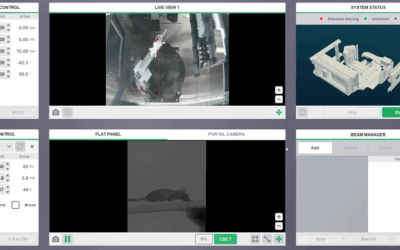
The Value of SARRP
Researchers used SARRP’s CBCT image guidance for irradiation. CBCT scans were performed for treatment planning, and dose volume histograms (DVHs) were extracted from Muriplan.