Xstrahl in Action
In radiology, low X-ray energies (<140 keV) are used to obtain an optimal image while in radiotherapy, higher X-ray energies (MeV) are used to eradicate tumor tissue. In radiation research, both these X-ray energies are being used to extrapolate in vitro research to clinical practice. However, the energy deposition of X-rays depends on their energy spectrum, which might lead to changes in biological response. This study compared the DNA damage response (DDR) in peripheral blood lymphocytes (PBLs) exposed to X-rays with varying beam quality, mean photon energy (MPE) and dose rate.
Publication
Peripheral blood lymphocytes differ in DNA damage response after exposure to X-rays with different physical properties
Authors
Simon Sioen, Louise D’Hondt, Fien Van Houte, Robin Demuynck, Klaus Bacher, Carlos De Wagter, Anne Vral, et al.

Key Findings
-
The induction of chromosomal damage and initial (30 min) DNA double-stranded breaks (DSBs) were significantly higher for 220 kV X-rays compared to 6 MV X-rays, while cell death induction was similar.
-
Specific cell death inhibitors for apoptosis, necroptosis and ferroptosis were not capable of blocking cell death after irradiation using low or high-energy X-rays.
-
Additional Cu filtration increased the MPE, which significantly decreased the amount of chromosomal damage and DSBs. Within the tested ranges no specific effects of dose rate variation were observed.

Figure 1 shows the DDR in PBLs influenced by the beam quality and MPE. This study reinforces the need for consideration and inclusion of all physical parameters in radiation-related studies.

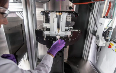
The Value of SARRP
Researchers used Xstrahl’s SARRP to irradiate cell cultures and determine if DDR in PBLs is influenced by the beam quality, MPE, and dose.