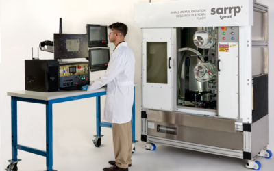
Bioluminescence imaging (BLI) is a non-contact, optical imaging technique based on measurement of emitted light due to an internal source, which is often directly related to cellular activity. It is widely used in pre-clinical small animal imaging studies (such as those preformed on the Xstrahl SARRP) to assess the progression of diseases such as cancer, aiding in the development of new treatments and therapies. For many applications, the quantitative assessment of accurate cellular activity and spatial distribution is desirable as it would enable direct monitoring for prognostic evaluation.
This requires quantitative spatially-resolved measurements of bioluminescence source strength inside the animal to be obtained from BLI images. This is the goal of bioluminescence tomography (BLT) in which a model of light propagation through tissue is combined with an optimization algorithm to reconstruct a map of the underlying source distribution.
As most models consider only the propagation of light from internal sources to the animal skin surface, an additional challenge is accounting for the light propagation from the skin to the optical detector. Existing approaches typically use a model of the imaging system optics (e.g. ray-tracing, analytical optical models) or approximate corrections derived from calibration measurements. However, these approaches are typically computationally intensive or of limited accuracy.
In the work “Quantitative bioluminescence tomography using spectral derivative data” Hamid Dehgani, James A Guggenheim, Shelly L Taylor, Xiangkun Xu and Ken Kang-hsin Wang, a new approach is presented in which, rather than directly using BLI images acquired at several wavelengths, the spectral derivative of that data (difference of BLI images at adjacent wavelengths) is used in BLT.
As light at similar wavelengths encounters a near-identical system response (path through the optics etc.) this eliminates the need for additional corrections or system models. This approach is applied to BLT with simulated and experimental phantom data and shown that the error in reconstructed source intensity is reduced from 49% to 4%. Qualitatively, the accuracy of source localization is improved in both simulated and experimental data, as compared to reconstruction using the standard approach.
This Xstrahl In Action was adapted from a article found on a National Library of Medicine website.