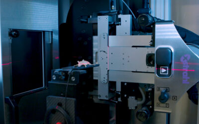
Although small animal image-guided radiotherapy (SA-IGRT) systems are used increasingly in preclinical research, tools for performing routine quality assurance have not been optimized and are not readily available. Robust, efficient, and reliable quality assurance tools are needed to ensure the accuracy and reproducibility of SA-IGRT systems. Several investigators have reported custom-made phantoms and protocols for SA-IGRT systems quality assurance. These are typically time and resource intensive and are therefore not well suited to the preclinical radiotherapy environment, in which physics support is limited and routine quality assurance is performed by technical staff.
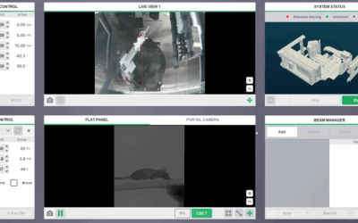
In their paper “Development and implementation of EPID-based quality assurance tests for the small animal radiation research platform (SARRP)” Anvari A, Poirier Y and Sawant A investigated the use of the inbuilt electronic portal imaging device (EPID) to develop and validate routine quality assurance tests and procedures. In their work, they focus on the Xstrahl Small Animal Radiation Research Platform (SARRP) EPID. However, the methodology and tests developed here are applicable to any SA-IGRT system that incorporates an EPID.
They performed a comprehensive characterization of the dosimetric properties of the camera-based EPID at kilovoltage energies over a 11-month period, including detector warm-up time, radiation dose history effect, stability and short- and long-term reproducibility, gantry angle dependency, output factor, and linearity of the EPID response. In response they developed a test to measure the constancy of beam quality in terms of half-value layer and tube peak potential using the EPID. We verified the SARRP daily output and beam profile constancy using the imager. We investigated the use of the imager to monitor beam-targeting accuracy at various gantry and couch angles.
They found that the EPID response was stable and reproducible, exhibiting maximum variations of ≤0.3% and ≤1.9% for short and long terms, respectively. The detector showed no dependence on response at different gantry angles, with a maximum variation ≤0.5%. They found close agreement in output factor measurement between the portal imager and reference dosimeters, with maximum differences ≤3% for ionization chamber and ≤1.7% for Gafchromic EBT3 dosimetry film, respectively. In doing so they have shown that the EPID response is linear with tube current (mA) for the entire range of tube kilovoltage peak. Notably, a close relationship was seen between the detector response vs mA slope, and the kilovoltage peak, allowing an independent verification of kilovoltage peak stability based solely on EPID response. In addition to dosimetry tests, according to the beam-targeting measurement using portal images, maximum displacement of the central axis of the x-ray beam (due to sag) was 0.76 ± 0.09 mm at gantry 135°/couch 0° and 0.89 ± 0.06 mm at gantry 0°/couch -135°.
The first comprehensive analysis on the dosimetric properties of an EPID operating at kilovoltage x-ray energies was performed in this study and as a result they characterized the detector performance over a 11-month period. The results indicate that the imager is a stable and convenient tool for SARRP routine quality assurance tests.
This Xstrahl In Action was adapted from a article found on a National Library of Medicine website.


