


Expanding Preclinical Translational Research into Precision Radiotherapy
REPLICATE CLINICAL PRACTICE
SECURE GRANT FUNDING

PERFORM SUCCESSFUL STUDIES
SARRP Replicates Modern Radiation Therapy Systems
SARRP enables investigators to perform clinically relevant radiation research in a preclinical setting. It is the only commercially available system that includes an integrated Motorized Variable Collimator (MVC) so researchers can perform conformal radiation therapy techniques commonly used in clinical practice. Dynamic jaws allow investigators to dictate field sizes ranging from 1mm x 1mm, up to 40mm x 80mm, so that it is easy to perform both small and large field irradiation. The 360° gantry and motorized stage allow for non-coplanar beam delivery from any angle, resulting in a more 3D conformal dose to the target area, while drastically minimizing normal tissue and surrounding tissue toxicity.
Advanced Treatment Planning Software
SARRP’s open system integration offers researchers flexibility to select from several treatment planning solutions to create and deliver simple or complex beam arrangements in a clinically relevant way. Techniques utilizing planar static beams, parallel opposed beams, continuous arc therapies, multiple isocenter treatments, and non-planar arcs can all be planned, evaluated, and delivered with SARRP. Even researchers with little to no knowledge of radiation therapy are able to easily plan and execute studies using either SmART-XPS or Muriplan.
SARRP Brings Success & Fosters Collaboration
SARRP is the first choice of researchers worldwide for translational radiation research. Diverse groups – including academic institutions, research hospitals, CROs, and countless interdisciplinary teams – conduct successful image-guided studies using SARRP. More than 85% of published studies are performed using SARRP. These investigators value integrated tools, such as:
- Motorized Variable Collimator
- Imaging Panel – 25cm x 20cm
- Monte Carlo Planning Algorithm
- Heated Mouse Bed
- 4D Treatment Head
WATCH OUR SARRP SERIES
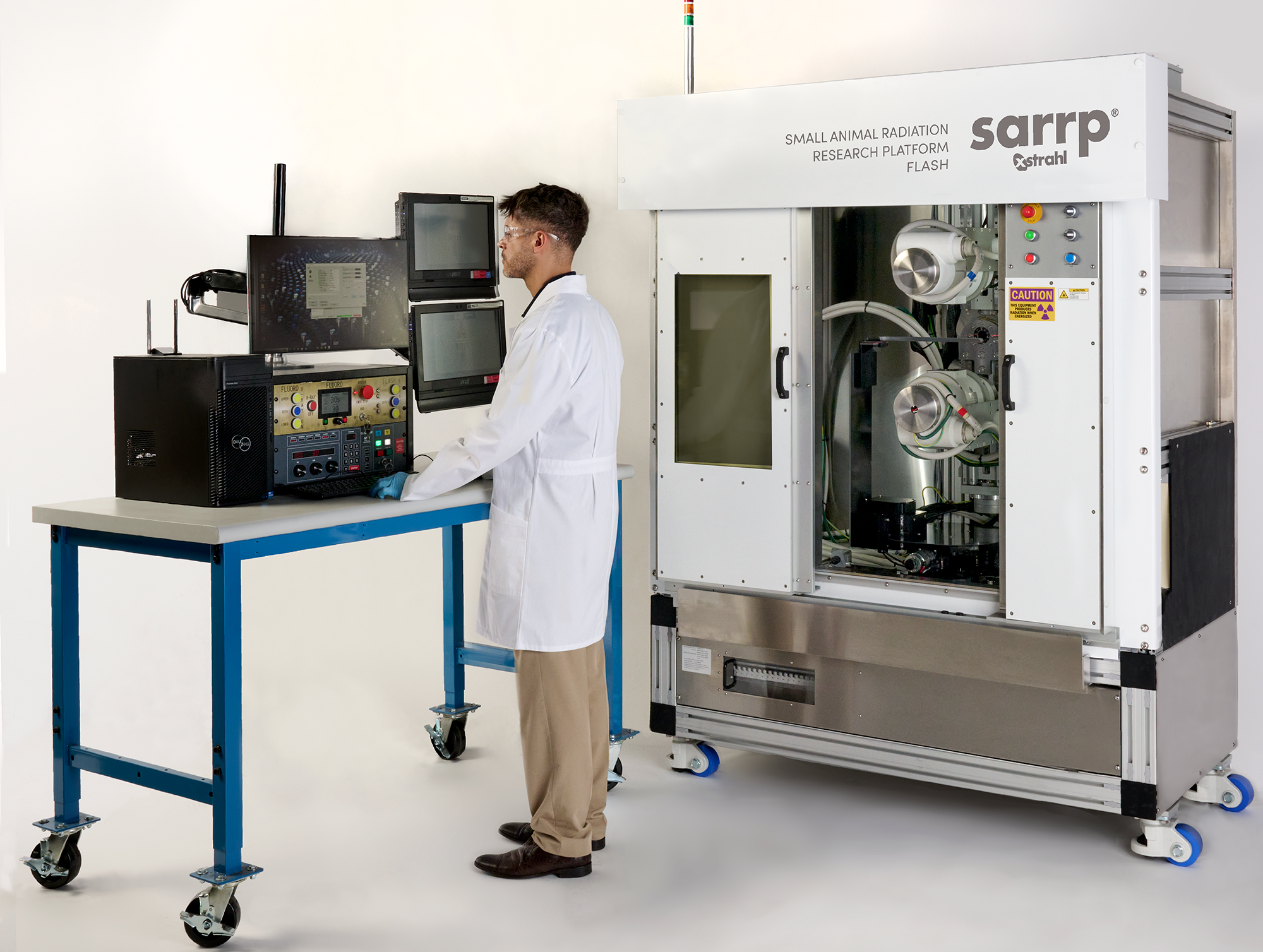
Small Animal Radiation Research Platform
SARRP is a customizable and powerful research platform designed to provide the preclinical research community with the ability to engage in the most clinically relevant, reproducible, and technically advanced studies.
REQUEST MORE INFORMATION
Contact us to learn more about SARRP and how Xstrahl can help you meet your research goals.

Community of Experts
Installed in nearly 100 facilities worldwide, SARRP has been cited as the primary research system employed in hundreds of peer-reviewed research publications, demonstrating the high quality of radiation research that can be performed using SARRP image-guided irradiation. A community has developed around the technology – connecting scientists, inspiring new studies, and advancing research around the world. The halo effect of bringing researchers together is an invaluable benefit of using SARRP.
RESOURCES
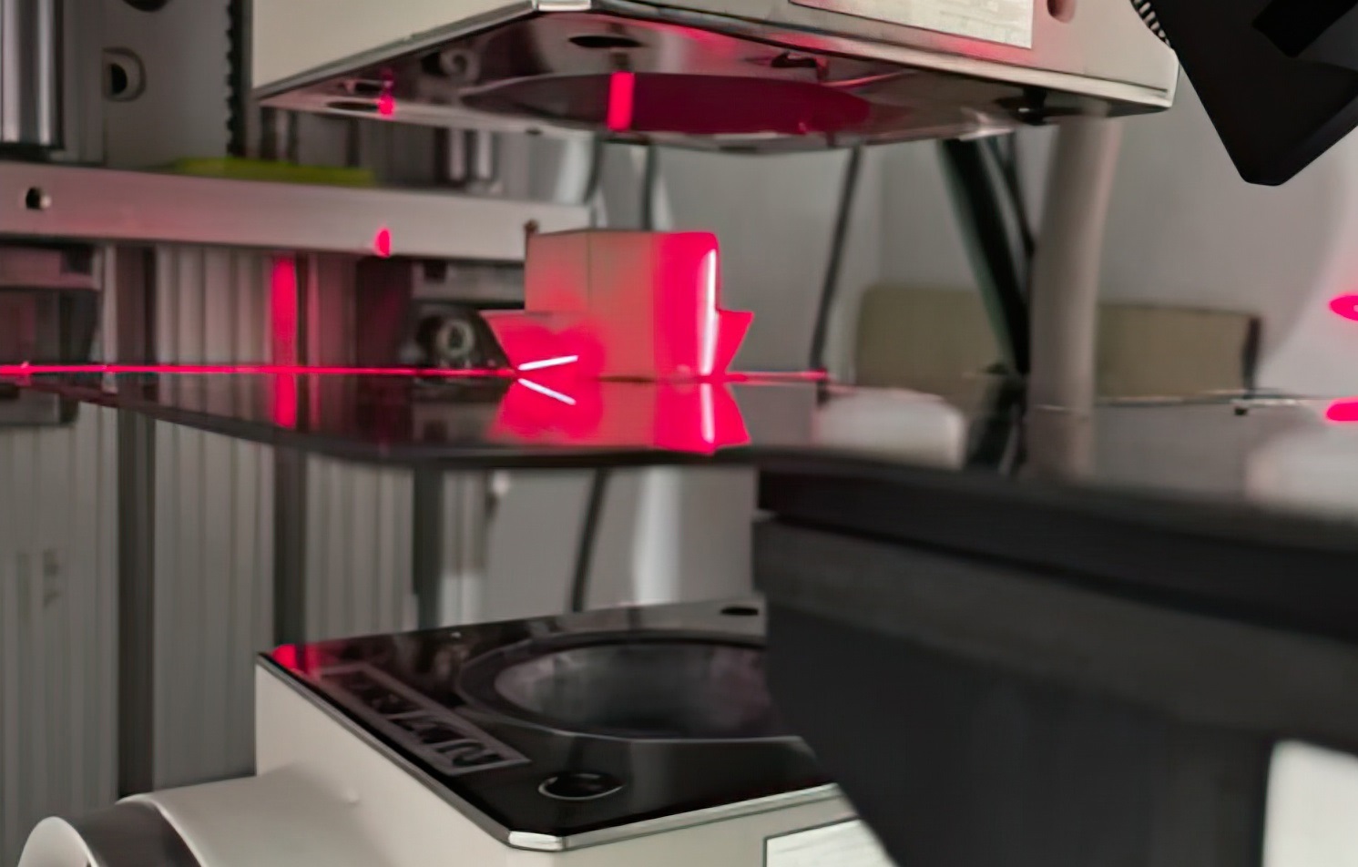

ADVANCED CAPABILITIES
SARRP enables deeper investigation and insight into how radiation interacts with tissue and the mechanisms of DNA repair. Clinical observations also underpin reverse translation studies.
Contact Us
Find out more about how Xstrahl will work tirelessly for you
Request More Information
Interested in Learning More?
Find out how you can implement cost-effective radiation treatment in your practice quickly and easily.