PURPOSE:
The goal of this work was to design a realistic mouse phantom as a useful tool for accurate dosimetry in radiobiology experiments.
METHODS:
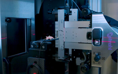
A subcutaneous tumor-bearing mouse was scanned in a microCT scanner, its organs manually segmented and contoured. The resulting geometries were converted into a stereolithographic file format (STL) and sent to a multi-material 3D printer. The phantom was split into two parts to allow for lung excavation and 3D-printed with an acrylic-like material and consisted of the main body (mass density ρ=1.18 g/cm3 ) and bone (mass density ρ=1.20 g/cm3 ). The excavated lungs were filled with polystyrene (ρ=0.32 g/cm3 ). Three cavities were excavated to allow the placement of a 1-mm diameter plastic scintillator dosimeter (PSD) in the brain, the center of the body and the subcutaneous tumor. Additionally, a laser-cut Gafchromic film can be placed in between the two phantom parts for 2D dosimetric evaluation. The expected differences in dose deposition between mouse tissues and the mouse phantom for a 220-kVp beam delivered by the small animal radiation research platform (SARRP) were calculated by Monte Carlo (MC).
RESULTS:
MicroCT scans of the phantom showed excellent material uniformity and confirmed the material densities given by the manufacturer. MC dose calculations revealed that the dose measured by tissue-equivalent dosimeters inserted in the phantom in the brain, center of the body, and the subcutaneous tumor would be underestimated by 3-5%, which is deemed to be an acceptable error assuming the required 5% accuracy of radiobiological experiments.
CONCLUSIONS:
The low-cost mouse phantom can be easily manufactured and, after a careful dosimetric characterization, may serve as a useful tool for dose verification in a range of radiobiology experiments. This article is protected by copyright. All rights reserved.
Esplen N, Alyaqoub E, Bazalova-Carter M.


