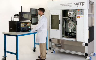
In their study “MicroCT imaging dose to mouse organs using a validated Monte Carlo model of the small animal radiation research platform (SARRP)” Johnstone CD and Bazalova-Carter M, establish imaging dose to mouse organs with a validated Monte Carlo (MC) model of the image-guided Small Animal Radiation Research Platform (SARRP) and to investigate the effect of scatter from the internal walls on animal therapy dose determination.
A MC model of the SARRP was built in the BEAMnrc code and validated with a series of homogeneous and heterogeneous phantom measurements. A segmented microCT scan of a mouse was used in DOSXYZnrc to determine mouse organ microCT imaging doses to 15-35 g mice for the SARRP pancake (mouse lying on couch) and standard (mouse standing on couch) imaging geometries for 40-80 kVp tube voltages. Imaging dose for off-center positioning shifts and maintaining image noise across tube voltages were also calculated. Half-value layer (HVL) measurements for the 220 kVp therapy beam in the presence of the SARRP shielding cabinet were modeled in BEAMnrc and compared to the 100 cm source-to-detector distance (SDD) in the scatter free, narrow-beam geometry recommended by the American Association of Physicists in Medicine Task Group 61 (AAPM TG-61).
For a 60 kVp, 0.8 mA, and 60 s scan protocol, maximum mean organ imaging doses to boney and non-boney structures were 10.5 cGy and 3.5 cGy, respectively, for an average size 20 g mouse. Current-exposure combinations above 323, 203, 147, 116, and 95 mAs for 40-80 kVp tube voltages, respectively, will increase body doses above 10 cGy. MicroCT mean body dose was 18% lower in pancake compared to standard imaging geometry. An 11% difference in measured HVL at a 50 cm SDD was found compared to MC simulated HVL for the AAPM TG-61 recommended scatter free geometry at a 100 cm SDD. This change in HVL resulted in a 0.5% change in absorbed dose to water calculations for the treatment beam.
This Xstrahl In Action was adapted from a article found on a National Library of Medicine website.


