PURPOSE:
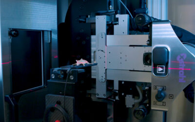
To characterize a small plastic scintillator developed for high resolution, real-time dosimetry of therapy and imaging x-ray beams delivered by an image-guided small animal irradiator.
MATERIALS AND METHODS:
A 1 mm diameter, 1 mm long polystyrene BCF-60 scintillating fiber dosimeter was characterized with 220 kVp therapy and 40, 50, 60, 70, and 80 kVp imaging beams on the Small Animal Research Platform (SARRP). Scintillator output, sensitivity (charge per unit dose), linearity, and 0.2-mm resolution beam profile measurements were performed. A validated in-house Monte Carlo (MC) model of the SARRP was used to compute detailed energy spectra at locations of dosimetry, and validated scintillator measurement with MC simulations. Mass energy-absorption coefficients from the National Institute of Standards and Technology (NIST) tables convolved with MC-derived spectra were used in conjunction with Birks ionization quenching factors to correct scintillator output. An air-kerma calibration method was employed to correct scintillator output for in-air beam profile measurements with open, 5 x 5, and 3 x 3 mm2 square field sizes, and compared to MC simulations.
RESULTS:
Scintillator dose response showed excellent linearity (R2 ≥ 0.999) for all sensitivity measurements, including output as a function of tube current. Detector sensitivity was 2.41 μC Gy-1 for the 220 kVp therapy beam, and it ranged from 1.21 to 1.32 μC Gy-1 for the 40-80 imaging beams. Percentage difference in sensitivity between the therapy and imaging beams before sensitivity correction and after using the Birks quenching factors were 52.3% and 10.2%, respectively. Percentage differences between the therapy and imaging beam sensitivities after using the air kerma calibration method for in-air measurements was excellent and below 0.3%. In-air beam profile measurements agreed to MC simulations within a mean difference of 2.4% for the 5 x 5 and 3 x 3 mm2 field sizes, however, the scintillator showed signs of volume averaging at the penumbra edges.
CONCLUSIONS:
A small plastic scintillator was characterized for therapy and imaging energies of a small animal irradiator, with output corrected for using an in-house MC model of the irradiator. The characterization of the scintillator detector system for small fields presents steps towards implementing real-time measurements for quality assurance and small animal treatment and imaging dose verification. This article is protected by copyright. All rights reserved.
Johnstone CD, Therriault-Proulx F, Beaulieu L, Bazalova-Carter M.


