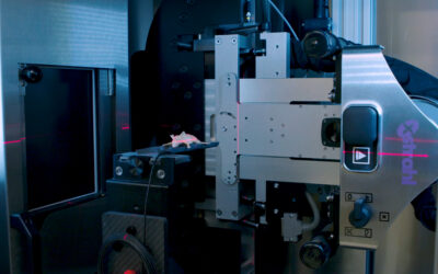
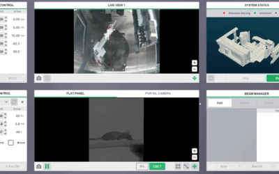
There are many unknowns in the radiobiology of proton beams and other particle beams. We describe the development and testing of an image-guided low-energy proton system optimized for radiobiological research applications. A 50 MeV proton beam from an existing cyclotron was modified to produce collimated beams (as small as 2 mm in diameter). Ionization chamber and radiochromic film measurements were performed and benchmarked with Monte Carlo simulations (TOPAS). The proton beam was aligned with a commercially-available CT image-guided x-ray irradiator device (SARRP, Xstrahl Inc.). To examine the alternative possibility of adapting a clinical proton therapy system, we performed Monte Carlo simulations of a range-shifted 100 MeV clinical beam. The proton beam exhibits a pristine Bragg Peak at a depth of 21 mm in water with a dose rate of 8.4 Gy min-1 (3 mm depth). The energy of the incident beam can be modulated to lower energies while preserving the Bragg peak. The LET was: 2.0 keV µm-1 (water surface), 16 keV µm-1 (Bragg peak), 27 keV µm-1 (10% peak dose). Alignment of the proton beam with the SARRP system isocenter was measured at 0.24 mm agreement. The width of the beam changes very little with depth. Monte Carlo-based calculations of dose using the CT image data set as input demonstrate in vivo use. Monte Carlo simulations of the modulated 100 MeV clinical proton beam show a significantly reduced Bragg peak. We demonstrate the feasibility of a proton beam integrated with a commercial x-ray image-guidance system for preclinical in vivo studies. To our knowledge this is the first description of an experimental image-guided proton beam for preclinical radiobiology research. It will enable in vivo investigations of radiobiological effects in proton beams.
Ford E, Emery R, Huff D, Narayanan M, Schwartz J, Cao N, Meyer J, Rengan R, Zeng J, Sandison G, Laramore G, Mayr N.


