The consequences of low to moderate doses of ionizing radiation (IR), especially for children exposed to CT scans, remains a challenging scientific topic. So far, no proper experimental models are available to better comprehend the consequences of exposure of the postnatal brain to IR, its effects on behavioral functions, and all related pathophysiological mechanisms.
Recently, in the publication of Dos Santos et al. entitled ‘Development of whole brain versus targeted dentate gyrus irradiation model to explain low to moderate doses of exposure effects in mice’, published in the journal of Scientific Reports, scientists have developed and described new tools to enhance scientific understanding on better protection of children exposed to IR.
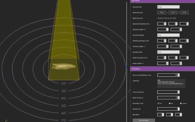
Three novel experimental models in 10-days-old mice were developed: one model of brain irradiation and two targeted irradiation models of the dorsal and ventral dentate gyrus. Using the small animal radiation research platform (SARRP), low to moderate doses of IR (less than 1Gy) were executed to the regions of interest. Coupling of the CT imaging build of SARRP and co-registration with MRI imaging enabled optimal localization yielding accurate treatment planning. Their irradiation strategy has been two-fold endorsed. The millimetric ballistic specificity of their mouse models was first validated by measuring gamma-H2AX increase after irradiation. As expected, increased gamma-H2AX staining was dose-dependent and acted as an irradiation dose readout. As such, the immuno-histochemical procedure deciphered high accuracy and reproducibility of their irradiation strategy. Next, higher anxiety/depressive-like behavior, preferentially mediated by the ventral part of the dentate gyrus, in mice after brain and ventral dentate gyrus IR exposure, has been demonstrated and described.
Scientists concluded that their findings enabled ballistic and functional validation of experimental irradiation models that can be used in parallel to (i) evaluate brain radiosensitivity, (ii) evaluate radiosensitivity of millimetric brain structures and to (iii) elucidate pathophysiological mechanisms involved in radiation-induced behavioral disorders, considering the functional brain’s organizational complexity. These three models are crucial to investigate radiation-induced cognitive disorders and the involvement of adult neurogenesis alterations of the dorsal and ventral DG hippocampus in their pathophysiology. Of note, the technological properties of SARRP facilities and parallel recording of MR and CBCT images favors visualization of structures of interest and enables high millimetric ballistic specificity.
This Xstrahl In Action was adapted from a article found on a National Library of Medicine website.