Despite both clinical and pre-clinical technological advancements, a lack of effective treatment for patients with primary brain tumors and those originating from metastasis remains. With the 5-year survival rate of patients experiencing primary glioblastoma multiforme less than 20%, and less than 10% of those with brain metastases originating from melanoma (Sandru et al., J Med Life. 2014; American Cancer Society). Translational pre-clinical models which yield clinically relevant results have never been more imperative. Pre-clinical models that replicate human disease are a necessity to drive the understanding of translational cancer biology forward. With clinical practice making technological advancements to more accurately treat patients (MRI+Linac). It now becomes a responsibility of biotechnology companies, and pre-clinical investigators to keep pace with clinical advancement.
Depending on the size and location of the primary or metastasized lesion, cone-beam computed tomography (CBCT) is normally combined with other imaging modalities like magnetic resonance (MR), or positron emission tomography (PET). These technologies enable increased accuracy of tumor identification, measurement, and treatment. Recently, a group of investigators from Columbia University lead by Dr. Simon K. Cheng have developed an orthotopic model of melanoma brain metastasis where they have utilized SARRPs image registration capabilities to accurately fuse T1 post contrast MRIs with the SARRPs 3D reconstructed CBCT image (Wu et al., IJROBP, 2017). Here, the investigators characterized stereotactic radiosurgery (SRS), tumor response, MR image fusion, and validation of SARRP dosimetry using a unique mouse-like phantom.
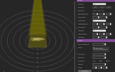
In an attempt to mimic clinical melanoma brain metastasis, B16 melanoma cells were injected orthotopically into the brain of C57BL/6 mice. Tumor-bearing mice were then imaged 11 days post injection, where tumors were contoured and then registered with SARRP CBCT for treatment consisting of a 60-degree sagittal arc to 18Gy using a 3x3mm collimator (Figure 1).
Furthermore, both axial and sagittal mouse-like phantom based radiochromic film analysis was performed (Figure 2 & 3). This was done as a measure of assurance to access radiation dose, distribution, and targeting accuracy. After analysis of the radiochromic film, it was observed that the maximum dose measured in both sagittal and axial phantoms are representative of the expected doses. Additionally, isodose lines in the axial phantom are similar to what was generated by SARRP. However, variability in the isodose lines measured in the sagittal phantom was observed. The authors suggested a limitation in the capability of the sagittal phantom to measure dose due to the film being sandwiched between the sagittal layers of the phantom.
The ability to fuse MR images with the SARRP cone beam-CT allows for not only increased targeting accuracy, but also the ability to visualize the tumor response. Here, the group at Columbia exploited this technique and showed that mice experiencing intracranial B16 melanoma tumors responded favorably to the SRS treatment, and had an increase in animal survival compared to those that did not receive SRS (Figure 4 & 5).
The authors conclude that “this is the first study to demonstrate accurate SRS delivery using the SARRP in a melanoma mouse intracranial model validated using a mouse-like phantom.” This study performed by the group led by Dr. Cheng truly showcases the dynamic capability of the SARRP and clinically relevant methods that push its functional capacity. Furthermore, the authors have focused on executing an experimental process that drives the understanding of cancer biology forward. This is something that’s imperative if we as a scientific community want to be more effective in clinical practice.
The full article can be accessed at http://www.redjournal.org/article/S0360-3016(17)30913-6/pdf.
Cheng-Chia Wu, Kunal R. Chaudhary, Yong Hum Na, David Welch, Paul J. Black, Adam M. Sonabend, Peter Canoll, Yvonne M. Saenger, Tony J.C. Wang, Cheng-Shie Wuu, Tom K. Hei, Simon K. Cheng. Quality assessment of stereotactic radiosurgery of a melanoma brain metastases model using a mouse-like phantom and the small animal radiation research platform. IJROBP. In Press. 2017.
To find out more about the science being conducted with Xstrahl systems you can visit Xstrahl’s website to learn more or Xstrahl Life Sciences for the latest developments.
This Xstrahl In Action was adapted from a article found on a National Library of Medicine website.