Introduction
Preclinical CT-guided radiotherapy platforms are increasingly used but the CT images are characterized by poor soft tissue contrast. The aim of this study was to develop a robust and accurate method of MRI-guided radiotherapy (MR-IGRT) delivery to abdominal targets in the mouse.
Methods
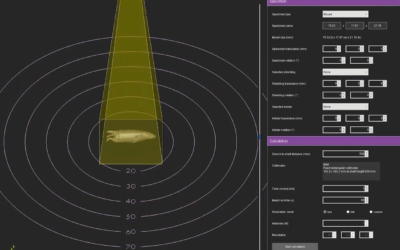
A multimodality cradle was developed for providing subject immobilisation and its performance was evaluated. Whilst CT was still used for dose calculations, target identification was based on MRI. Each step of the radiotherapy planning procedure was validated initially in vitro using BANG gel dosimeters. Subsequently, MR-IGRT of normal adrenal glands with a size-matched collimated beam was performed. Additionally, the SK-N-SH neuroblastoma xenograft model and the transgenic KPC model of pancreatic ductal adenocarcinoma were used to demonstrate the applicability of our methods for the accurate delivery of radiation to CT-invisible abdominal tumours.
Results
ConclusionThis is, to our knowledge, the first study to demonstrate preclinical MR-IGRT in intra-abdominal organs. The proposed MR-IGRT method presents a state-of-the-art solution to enabling robust, accurate and efficient targeting of extracranial organs in the mouse and can operate with a sufficiently high throughput to allow fractionated treatments to be given.
Veerle Kersemans, John S. Beech, Stuart Gilchrist, Paul Kinchesh, Philip D. Allen, James Thompson, Ana L. Gomes, Zenobia D’Costa, Luke Bird, Iain D. C. Tullis, Robert G. Newman, Aurelien Corroyer-Dulmont, Nadia Falzone, Sean C. Smart